X-Ray and Imaging Technology
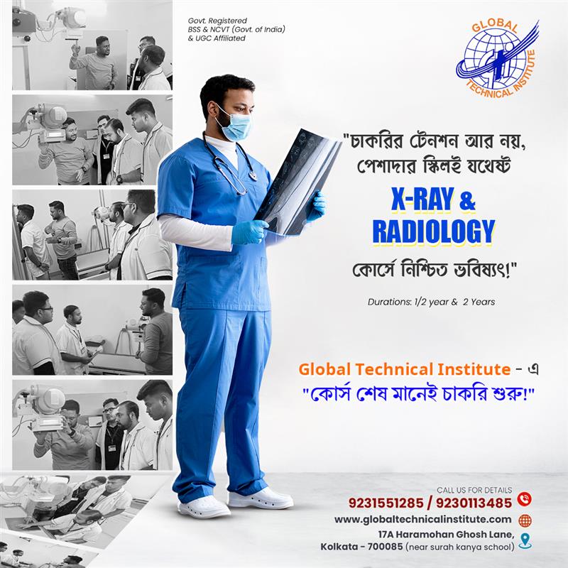
Choosing the right healthcare training course is an important decision. If you are interested in medical technology, patient care and diagnostic services, X-Ray and Imaging Technology can be a career option worth exploring.
Today, medical imaging plays an important role in modern healthcare. Doctors use X-rays and other imaging tests to examine bones, joints, the chest and different parts of the body. These investigations help medical professionals identify injuries, detect health conditions and plan further treatment.
However, working in this field requires more than knowing how to operate an X-ray machine. A trained imaging professional must understand patient positioning, radiation protection, equipment handling, image quality, infection control and clinical safety.
If you are searching for an X-Ray Training Institute in Kolkata, this guide will help you understand the course options, eligibility, duration, syllabus, practical training, fees, admission process, career opportunities and important points to check before enrolling.
At Global Technical Institute (GTI), we understand that students need clear information before choosing a professional course. Therefore, we have explained the subject from the basics and included the details published on our X-Ray & Imaging Technology course page.
1. What Is X-Ray and Imaging Technology?
X-Ray and Imaging Technology is a branch of healthcare technology that uses imaging equipment to create pictures of structures inside the human body.
An X-ray machine produces a controlled amount of X-ray radiation. When the radiation passes through the body, different tissues absorb different amounts of it. A detector captures the remaining radiation and produces an image.
Doctors use these images to examine different parts of the body.
For example:
- Chest X-rays help doctors examine the lungs and chest structures.
- Bone X-rays help identify fractures and certain bone conditions.
- Dental X-rays help examine teeth and surrounding structures.
- Joint X-rays help assess certain bone and joint problems.
- Abdominal X-rays may help doctors investigate selected abdominal conditions.
X-ray imaging is one part of the wider field of radiology and medical imaging. Other imaging technologies include computed tomography (CT), magnetic resonance imaging (MRI), ultrasound and mammography.
A trained radiography professional works with imaging equipment and supports the diagnostic process under the applicable clinical supervision and professional responsibilities.
2. What Does an X-Ray Technician Do?
An X-ray technician, also known as a radiographer or radiology technologist depending on the qualification and role, helps produce medical images for diagnostic purposes.
The exact duties depend on the qualification, workplace, equipment and level of responsibility.
Common responsibilities may include:
2.1 Preparing the X-Ray Room
Before an examination, the professional checks whether the imaging room is ready.
This may involve:
- Checking the X-ray equipment.
- Checking the examination table and accessories.
- Keeping positioning aids ready.
- Maintaining a clean examination area.
- Following equipment safety procedures.
- Confirming that required protective measures are available.
2.2 Identifying and Preparing the Patient
Patient identification is an important part of imaging safety.
The professional may need to:
- Confirm the patient’s identity using the approved process.
- Check the requested examination.
- Explain the basic procedure.
- Ask relevant safety-screening questions.
- Help the patient prepare for the examination.
- Maintain patient privacy and dignity.
The correct examination must be confirmed before proceeding.
2.3 Patient Positioning
Correct positioning is essential for producing a useful X-ray image.
The patient may need to stand, sit or lie down depending on the examination. The professional positions the body part according to the prescribed imaging protocol.
Incorrect positioning can lead to poor image quality and may require another exposure. This is why positioning is an important part of radiography training.
2.4 Operating Imaging Equipment
An appropriately qualified professional operates the equipment within their authorised scope of practice.
This involves understanding:
- Equipment controls.
- Exposure factors.
- Image receptors.
- Examination protocols.
- Safety interlocks.
- Basic equipment checks.
Equipment operation must always follow the manufacturer’s instructions, institutional protocols and radiation-safety requirements.
2.5 Checking Image Quality
After an exposure, the image is checked for technical quality.
Important points include:
- Correct positioning.
- Appropriate image coverage.
- Suitable exposure.
- Sharpness.
- Visibility of the required anatomical structures.
- Avoidance of unnecessary repeat exposures.
The radiologist or authorised medical professional interprets the images and prepares the diagnostic report. An X-ray technician must not independently diagnose a patient or provide a medical report unless separately qualified and legally authorised to do so.
2.6 Maintaining Patient and Radiation Safety
Safety is one of the most important responsibilities in medical imaging.
Professionals must follow radiation-protection procedures, use approved protective measures where applicable, and avoid unnecessary radiation exposure.
They must also maintain patient records and follow the healthcare facility’s infection-control procedures.
3. Why Choose X-Ray Technician Training in Kolkata?
Kolkata has a large healthcare network that includes government hospitals, private hospitals, nursing homes, diagnostic centres and specialised medical facilities.
These facilities use different forms of medical imaging to support diagnosis and treatment.
Students interested in healthcare technology can explore radiography as a professional learning pathway.
Some reasons students consider this field include:
1. Healthcare-related career: The work supports doctors and other healthcare professionals in diagnostic services.
2. Technical learning: Students develop knowledge of imaging equipment, radiation physics and image production.
3. Patient interaction: The role involves communicating with patients and helping them prepare for examinations.
4. Different workplace options: Qualified professionals may find opportunities in hospitals, diagnostic centres and other healthcare facilities, depending on their qualification and eligibility.
5. Scope for further learning: Students may explore advanced imaging education after completing the required recognised qualification.
However, students should remember that employment depends on the qualification, practical competence, registration requirements where applicable, employer criteria and available vacancies. Completing a course does not guarantee a job.
4. Types of X-Ray and Radiology Training Courses
Before joining an institute, it is important to understand the difference between a certificate course, a diploma and a degree.
These qualifications are not automatically equivalent. Their recognition, duration, eligibility and professional value depend on the awarding body and applicable regulations.
4.1 Certificate Course in X-Ray Technology
A certificate course generally introduces students to basic concepts of X-ray imaging.
Depending on the course structure, subjects may include:
- Introduction to medical imaging.
- Basic radiation physics.
- X-ray equipment awareness.
- Patient preparation.
- Basic positioning concepts.
- Radiation-safety awareness.
- Image-quality basics.
- Healthcare communication.
A certificate course may be suitable for students who want introductory knowledge or a basic understanding of the subject.
However, a short-term certificate should not be assumed to qualify someone for independent professional practice as a registered radiographer or imaging technologist.
Before admission, students should ask whether the certificate is recognised for the job role they want to pursue.
4.2 Diploma in X-Ray Technology
A diploma course generally provides more extensive theoretical and practical learning than a short certificate programme.
A properly recognised radiography diploma may include:
- Human anatomy and physiology.
- Radiation physics.
- Radiographic techniques.
- Patient positioning.
- Imaging equipment.
- Radiation protection.
- Image processing.
- Clinical procedures.
- Patient care.
- Clinical internship or supervised practical training.
The duration and eligibility depend on the specific qualification and awarding authority.
For example, the State Medical Faculty of West Bengal lists a Diploma in Radiography (Diagnostic), with a published duration of two years plus six months of compulsory post-examination training.
The NCAHP’s 2026 competency-based D.MRIT framework also describes a two-year programme with a six-month clinical internship. It specifies Class 12 Science with Physics, Chemistry and Biology as the entry qualification.
These are formal qualification pathways. Students should verify the institution’s approval and the exact awarding authority before enrolling.
4.3 Bachelor’s Degree in Medical Radiology and Imaging Technology
A bachelor’s degree provides a broader academic and professional foundation in medical radiology and imaging.
Depending on the approved programme, subjects may include:
- Human anatomy.
- Human physiology.
- Radiation physics.
- General radiography.
- Imaging technology.
- CT imaging.
- MRI principles.
- Patient care.
- Radiation protection.
- Clinical practice.
- Research and professional ethics.
The degree duration and eligibility must be checked against the current university and NCAHP framework.
Do not assume that every institute offering a course with the words “B.Sc. Radiology” provides a recognised degree. Verify the university, programme approval, admission eligibility and registration pathway.
4.4 Certificate vs Diploma vs Degree: What Is the Difference?
| Course type | Main purpose | What students should verify |
| Certificate | Introductory or skill-based learning | Awarding body, course scope and accepted job roles |
| Diploma | Structured professional education | Government approval, recognised curriculum, duration and internship |
| Bachelor’s degree | Higher-level academic and professional education | Recognised university, approved programme and registration eligibility |
Important: The word “diploma” alone does not prove government recognition. Always verify the qualification and awarding authority.
5. X-Ray Course Eligibility: Who Can Apply?
Eligibility depends on the course and the authority awarding the qualification.
Students should not assume that the entry requirements for a private certificate programme are the same as those for a recognised radiography diploma.
5.1 Eligibility for a Basic Certificate Course
Some private institutes may offer introductory certificate programmes to students with different educational backgrounds.
However, students must check:
- Minimum educational qualification.
- Whether Science subjects are required.
- Age requirements, if any.
- Whether the certificate has an accepted professional purpose.
- Whether further recognised education is possible after completion.
5.2 Eligibility for a Recognised D.MRIT Course
The NCAHP’s 2026 D.MRIT framework specifies:
- Class 12 or equivalent from a recognised board.
- Science stream.
- Physics.
- Chemistry.
- Biology.
Students should check the latest admission notification and the approved institution’s requirements before applying.
5.3 Eligibility for a Bachelor’s Degree
A recognised bachelor’s programme generally requires Class 12 Science with the subjects and marks specified by the university and applicable regulations.
Admission may be based on merit, an entrance examination or another approved process.
5.4 Can Students Apply After Class 10?
Students who have completed Class 10 may find introductory or skill-based courses offered by private institutes.
However, Class 10 eligibility for a private certificate does not mean that the student meets the entry requirements for a recognised D.MRIT or bachelor’s degree.
If your long-term goal is to work as a qualified radiography professional, check the recognised qualification pathway first.
5.5 Is Biology Compulsory?
For the NCAHP D.MRIT pathway, Class 12 Science with Physics, Chemistry and Biology is specified.
Other courses may have different eligibility conditions. Always check the exact programme rather than relying on general statements about radiology admission.
6. X-Ray Course Duration in Kolkata
Course duration varies according to the qualification and awarding authority.
Here is a useful comparison:
| Programme | Published duration or structure |
| GTI Certificate in X-Ray & Imaging Technology | 1 year, as listed on the course page |
| GTI Diploma in X-Ray & Imaging Technology | 2 years, as listed on the course page |
| SMFWB Diploma in Radiography (Diagnostic) | 2 years + 6 months compulsory post-examination training |
| NCAHP D.MRIT framework | 2 years, including a six-month clinical internship |
The GTI course page lists a one-year certificate and a two-year diploma. It also lists two classes per week.
The SMFWB and NCAHP programmes are separate formal qualification pathways. They should not be treated as equivalent to GTI’s privately advertised course unless the required approval and recognition are established.
Students should ask the institute for the exact academic calendar, class hours, practical hours, examination schedule and completion requirements.
7. X-Ray Technician Course Syllabus: What Will You Learn?
A good X-Ray and Imaging Technology programme should cover both theoretical knowledge and practical skills.
The exact syllabus depends on the course and awarding body. The following subjects explain the main areas students should understand.
7.1 Human Anatomy
Anatomy is the study of the structure of the human body.
Radiography students need to understand the location and structure of bones, organs and body systems.
Important areas include:
- Skull and facial bones.
- Spine.
- Chest and ribs.
- Upper limbs.
- Lower limbs.
- Pelvis.
- Basic organ systems.
Anatomy helps students understand which body part is being examined and how it should be positioned.
7.2 Human Physiology
Physiology explains how the body functions.
Students learn the basic functions of the respiratory, circulatory, digestive, skeletal and other body systems.
This knowledge helps them understand the clinical purpose of different examinations.
7.3 Radiation Physics
Radiation physics introduces the principles behind X-ray production and image formation.
Topics may include:
- Basic atomic structure.
- Electromagnetic radiation.
- X-ray production.
- X-ray tube components.
- Radiation interaction with matter.
- Attenuation.
- Exposure factors.
- Image formation.
7.4 X-Ray Equipment and Components
Students should understand the purpose of major equipment components.
These may include:
- X-ray tube.
- Control panel.
- High-voltage generator.
- Collimator.
- Image detector.
- Examination table.
- Wall stand.
- Protective barriers.
- Digital image-processing system.
Understanding the equipment helps students follow operating procedures and identify basic technical issues.
7.5 Radiographic Positioning
Positioning is a core skill in radiography.
Students learn how the patient and body part are positioned for different examinations.
Examples include:
- Chest positioning.
- Skull positioning.
- Cervical spine positioning.
- Lumbar spine positioning.
- Shoulder positioning.
- Elbow positioning.
- Wrist positioning.
- Hand positioning.
- Pelvis positioning.
- Knee positioning.
- Ankle positioning.
- Foot positioning.
The actual procedure must follow the examination request and approved protocol.
7.6 Image Processing and Quality
Students learn the basic factors that affect image quality.
These include:
- Exposure.
- Contrast.
- Brightness.
- Sharpness.
- Noise.
- Positioning.
- Motion.
- Image artefacts.
In digital radiography, students may also learn about image receptors, digital processing and image storage systems.
7.7 Radiation Protection
Radiation protection is essential for patients, healthcare workers and the public.
Students should learn the principles of justification, optimisation and dose limitation.
They should understand:
- Why unnecessary exposure must be avoided.
- How collimation helps limit the exposed area.
- Why correct positioning matters.
- How protective barriers are used.
- The importance of controlled access to imaging rooms.
- The role of personal monitoring where required.
- The importance of following institutional safety protocols.
7.8 Patient Care and Communication
An imaging professional must communicate clearly and respectfully.
Students should learn how to:
- Explain the basic procedure.
- Guide patients through positioning.
- Support patients with limited mobility.
- Maintain privacy.
- Communicate with children and elderly patients.
- Follow patient-identification procedures.
- Recognise when assistance is needed.
- Escalate concerns to the appropriate healthcare professional.
7.9 Introduction to Advanced Imaging
A broader imaging programme may introduce students to other technologies.
These include:
- CT scan.
- MRI.
- Ultrasound.
- Mammography.
- Fluoroscopy.
- Digital radiography.
An introductory module does not automatically authorise a student to operate all these machines. Each technology requires appropriate education, supervised competency and professional authorisation.
8. Practical Training in X-Ray Technology
Practical learning is one of the most important parts of radiography education.
Students need more than classroom knowledge. They must understand how imaging procedures work in a controlled clinical environment.
A practical training programme may include the following areas, depending on the approved course structure.
8.1 Equipment Familiarisation
Students learn the names, functions and basic safety requirements of imaging equipment.
8.2 Positioning Demonstrations
Students observe and learn the correct positioning principles for different body parts.
8.3 Image Quality Assessment
Students learn how to identify common image-quality problems and understand why they occur.
8.4 Radiation Safety Practice
Students learn how to follow safety procedures and protect patients and staff from unnecessary exposure.
8.5 Patient Communication
Students practise explaining procedures and helping patients understand examination instructions.
8.6 Supervised Clinical Exposure
Recognised professional programmes include structured clinical learning.
The NCAHP D.MRIT framework includes a six-month clinical internship. Students should confirm the clinical training arrangements, location, supervision and completion documentation before admission.
Questions to ask an institute about practical training:
- How many practical hours are included?
- Is practical training conducted on actual imaging equipment?
- Is there a hospital or diagnostic-centre training arrangement?
- Who supervises students?
- Is the clinical training compulsory?
- How is practical competency assessed?
- Will students receive a documented internship or practical-training certificate?
Do not assume that a course includes hospital internship simply because its advertisement mentions practical learning.
9. X-Ray Equipment and Technologies Students Should Know
Medical imaging uses different types of equipment. Students should understand the basic purpose of each technology.
9.1 Conventional X-Ray Machine
A conventional X-ray machine produces images using X-ray radiation and an image receptor.
It is commonly used for chest, bone and other routine examinations.
9.2 Digital Radiography (DR)
Digital radiography uses electronic detectors to capture X-ray images.
The images can be processed and stored digitally.
9.3 Computed Radiography (CR)
Computed radiography uses imaging plates that are processed through a reader to produce digital images.
9.4 Portable X-Ray Machine
Portable X-ray equipment allows examinations to be performed near the patient when transferring them to a fixed imaging room is difficult.
It is commonly used in selected hospital settings.
9.5 CT Scanner
A CT scanner uses X-rays from multiple angles to create cross-sectional images.
CT is different from a routine X-ray examination and requires specific training.
9.6 MRI Scanner
MRI uses a strong magnetic field and radiofrequency signals to create images. It does not use ionising X-ray radiation.
MRI safety requires particular attention because of the magnetic field and associated equipment risks.
9.7 Ultrasound Equipment
Ultrasound uses high-frequency sound waves to create images. It does not use ionising radiation.
The use of ultrasound equipment requires the appropriate professional qualification and authorisation.
10. Radiation Safety: What Every Student Must Understand
Radiation safety is not an optional subject. It is a basic responsibility in medical imaging.
X-rays are a form of ionising radiation. Unnecessary exposure should be avoided.
Students should understand the following principles.
10.1 Justification
An examination should have a valid clinical reason. Imaging should not be performed without appropriate authorisation.
10.2 Optimisation
Radiation exposure should be kept as low as reasonably achievable while producing an image suitable for the clinical purpose.
10.3 Time, Distance and Shielding
These are basic radiation-protection concepts.
- Time: Avoid unnecessary time near a radiation source during exposure.
- Distance: Increasing distance from the source can reduce exposure.
- Shielding: Use appropriate protective barriers and equipment.
10.4 Pregnancy Screening
Pregnancy-related screening must follow the healthcare facility’s approved procedure.
If a patient may be pregnant, the concern should be referred to the authorised medical professional before proceeding.
10.5 Controlled Area
X-ray rooms must follow applicable safety requirements, access controls and warning-sign procedures.
10.6 Personal Monitoring
Where required, staff use personal monitoring devices to help assess occupational radiation exposure.
10.7 Equipment Quality Assurance
Imaging equipment must be maintained and tested according to applicable standards and institutional procedures.
Students should learn that safe imaging depends on trained staff, suitable equipment, correct protocols and proper supervision.
11. X-Ray Technician Admission Process in Kolkata
The admission process depends on the institute and the course.
Students can generally follow these steps.
Step 1: Choose the Course
Decide whether you want an introductory certificate, a recognised diploma or a bachelor’s degree.
Step 2: Check Eligibility
Confirm your educational qualification and required subjects.
Step 3: Verify the Institute
Check the awarding body, recognition, affiliation and approval documents.
Step 4: Understand the Course Structure
Ask for the syllabus, duration, class schedule, practical hours and examination details.
Step 5: Confirm the Fee
Request a complete written fee structure.
Step 6: Submit the Application
Complete the admission form and submit the required documents.
Step 7: Receive Admission Confirmation
Collect the admission receipt, course details, fee terms and academic schedule.
Step 8: Attend Classes and Practical Sessions
Follow the attendance, examination and practical-training requirements.
Step 9: Complete the Course
Meet the academic and assessment requirements.
Step 10: Collect Course Documents
Obtain the final certificate or diploma and supporting documents. For a recognised professional course, check the required registration process separately.
12. Documents Required for X-Ray Course Admission
The required documents depend on the institute and qualification.
Commonly requested documents may include:
- Recent passport-size photographs.
- Class 10 marksheet.
- Class 12 marksheet.
- School leaving or transfer certificate, if required.
- Identity proof.
- Address proof.
- Caste certificate, if applicable.
- Other documents specified by the admission authority.
Documents Listed by Global Technical Institute
The GTI X-Ray & Imaging Technology course page lists the following documents:
- Six copies of a photograph.
- Attested copies of the last passed marksheet.
- Madhyamik admit card.
- Any photo identity proof.
Students should confirm the current document requirements with the admissions team before submitting their application.
13. X-Ray Technician Course Fees in Kolkata
Course fees are an important factor when choosing an institute.
However, there is no single fee that applies to every X-Ray course in Kolkata. Fees can vary according to the qualification, duration, awarding body, practical facilities and clinical training arrangements.
The following factors may affect the total cost:
- Course type.
- Course duration.
- Admission fee.
- Tuition fee.
- Examination fee.
- Practical or laboratory fee.
- Clinical training charges.
- Study materials.
- Uniform or equipment charges.
- Certificate or diploma charges.
- Registration-related expenses, where applicable.
What Is the Fee at Global Technical Institute?
The published GTI X-Ray & Imaging Technology course page does not provide a specific fee amount.
Therefore, students should contact the institute for the latest certificate and diploma fee structure.
Questions to Ask Before Paying the Fee
Before admission, ask:
- What is the total course fee?
- Is the admission fee separate?
- Are examination charges included?
- Are practical-training charges included?
- Is there any additional internship fee?
- Is the fee refundable?
- Can the fee be paid in instalments?
- Will an official payment receipt be provided?
- Are there any charges for the final certificate?
- What happens if a student misses an examination?
Always request the complete fee structure in writing. Do not compare institutes only on the basis of the lowest advertised fee.
14. What Is the Salary of an X-Ray Technician in Kolkata?
Salary depends on several factors. There is no fixed salary for every person who completes an X-Ray course.
Important factors include:
- Type of qualification.
- Recognition of the qualification.
- Practical experience.
- Clinical competency.
- Employer requirements.
- Location.
- Hospital or diagnostic-centre size.
- Working hours.
- Job responsibilities.
- Additional imaging skills.
A newly qualified candidate may receive a different salary from an experienced radiographer.
Candidates with recognised qualifications, relevant clinical exposure and strong technical skills may be eligible for a wider range of roles.
How Can Students Improve Their Career Prospects?
Students can focus on:
- Completing an appropriate recognised qualification.
- Building strong anatomy knowledge.
- Learning correct positioning.
- Understanding radiation protection.
- Improving patient communication.
- Developing digital imaging knowledge.
- Completing supervised clinical training.
- Keeping their professional documents updated.
- Applying for roles that match their qualification.
Salary figures should be checked against current job advertisements and actual employer offers. An institute should not promise a fixed salary or guaranteed employment without verifiable evidence.
15. Career Opportunities After X-Ray Technician Training
Career options depend on the qualification, professional registration requirements and employer criteria.
People with appropriate recognised qualifications may explore roles in the following settings.
15.1 Hospitals
Hospitals use imaging services in emergency departments, outpatient departments, inpatient care and specialised units.
15.2 Diagnostic Centres
Diagnostic centres provide imaging services such as routine X-rays and other investigations.
15.3 Nursing Homes
Some nursing homes operate imaging facilities or work with external diagnostic service providers.
15.4 Specialised Imaging Centres
Qualified professionals may work in facilities that provide specialised imaging services, depending on their training and competence.
15.5 Government Healthcare Facilities
Government employment depends on the recruitment notification, prescribed qualification, registration requirements and selection process.
A private institute certificate does not automatically make a candidate eligible for a government radiographer post.
15.6 Further Education
Students may explore higher studies or advanced imaging education after completing the required recognised qualification.
The available progression depends on the awarding body and admission rules.
16. Skills Required to Become a Good X-Ray Technician
Technical knowledge is important, but a successful imaging professional also needs good communication and patient-care skills.
Technical Skills
- Understanding of anatomy.
- Knowledge of radiation physics.
- Patient positioning.
- Equipment awareness.
- Image-quality assessment.
- Radiation protection.
- Digital imaging basics.
- Clinical documentation.
Personal Skills
- Clear communication.
- Patience.
- Attention to detail.
- Professional behaviour.
- Teamwork.
- Time management.
- Problem-solving.
- Respect for patient privacy.
Patient-Care Skills
- Helping patients understand procedures.
- Supporting elderly patients.
- Communicating with children.
- Maintaining dignity.
- Recognising discomfort.
- Following infection-control practices.
- Escalating clinical concerns.
These skills help professionals work responsibly in a healthcare environment.
17. How to Choose the Best X-Ray Training Institute in Kolkata
Choosing an institute requires careful checking. A good advertisement alone is not enough.
Before admission, compare the following points.
17.1 Verify Recognition and Affiliation
Ask which organisation awards the certificate or diploma.
If the institute claims government recognition, request the relevant approval or affiliation document.
Check whether the approval applies to the exact course, campus and academic session.
17.2 Check the Course Duration
Confirm the exact duration and whether the stated period includes practical training or internship.
17.3 Review the Syllabus
The syllabus should explain the subjects, learning outcomes and assessment process.
17.4 Ask About Practical Facilities
Ask whether students receive supervised training on relevant equipment.
17.5 Confirm Clinical Training
If hospital or diagnostic-centre training is advertised, ask for the name of the training facility and written details of the arrangement.
17.6 Understand the Faculty
Ask about the qualifications and relevant professional experience of the faculty members.
17.7 Check the Fee Structure
Obtain a written breakdown of all fees.
17.8 Ask About Placement Support
If placement assistance is advertised, ask what it includes.
For example:
- Is there a placement cell?
- Are interviews arranged?
- Are employers identified?
- Is placement support available to every student?
- Are placement records available for verification?
Placement assistance is not the same as a guaranteed job.
17.9 Understand the Professional Scope
Ask whether the qualification supports registration or eligibility for the job you want.
17.10 Speak With the Admissions Team
Get your questions answered before paying the admission fee.
A responsible institute should explain the course structure and its limitations clearly.
18. X-Ray and Imaging Technology Course at Global Technical Institute, Kolkata
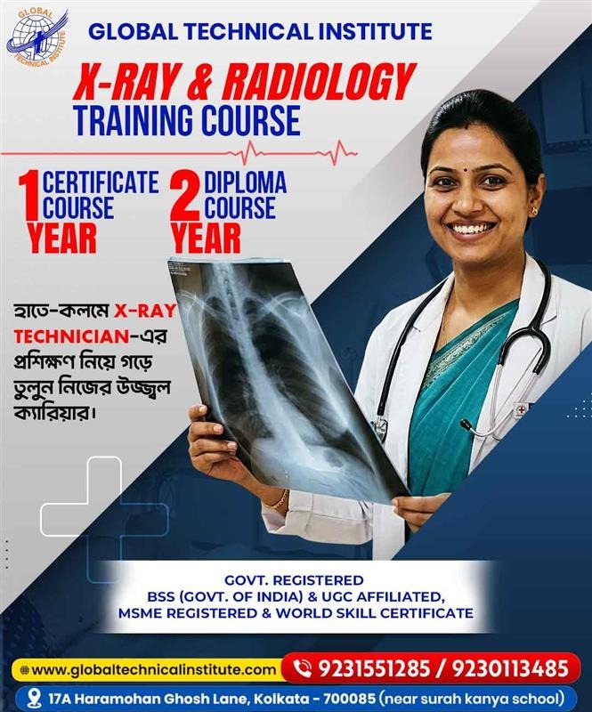
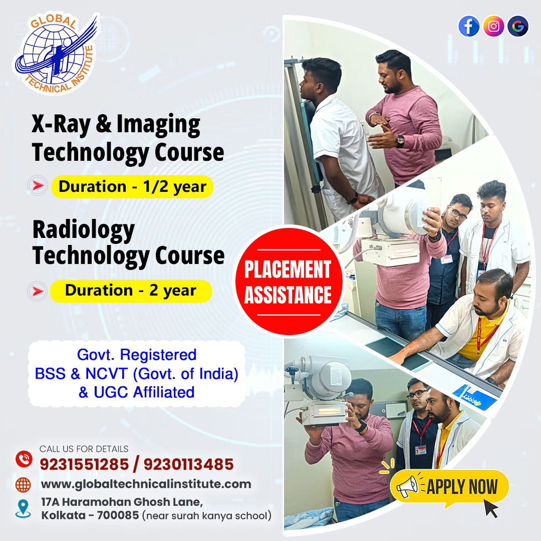
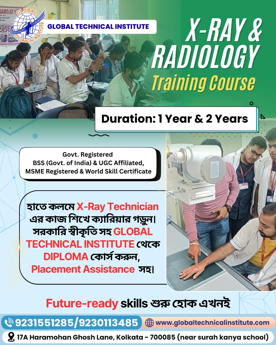
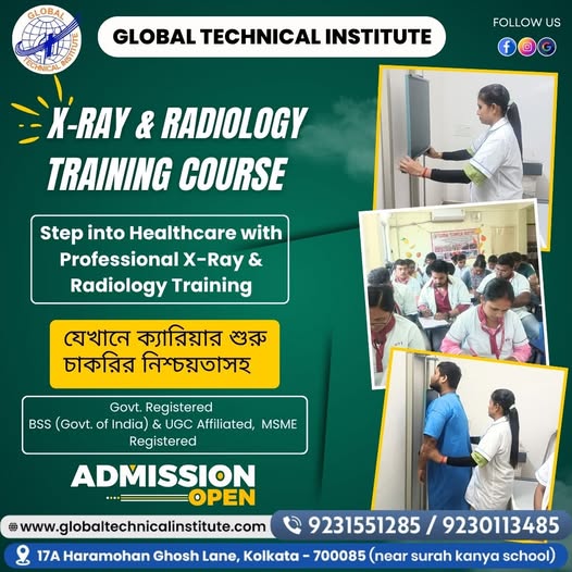
At Global Technical Institute (GTI), we offer X-Ray & Imaging Technology training for students interested in learning the fundamentals of medical imaging.
Our published course page lists two programme options:
- Certificate in X-Ray & Imaging Technology.
- Diploma in X-Ray & Imaging Technology.
Course Details Published by GTI
| Particular | Published information |
| Course name | X-Ray & Imaging Technology |
| Certificate option | Certificate in X-Ray & Imaging Technology |
| Diploma option | Diploma in X-Ray & Imaging Technology |
| Certificate duration | 1 year |
| Diploma duration | 2 years |
| Classes | 2 classes per week |
| Admission sessions | April, August and December |
| Minimum qualification | Higher Secondary pass |
| Skill-development module | Personality development and grooming |
| Board information | The page states “BSS Promoted by Govt. of India” |
| Institute certification | The page describes GTI as ISO certified |
The course page also describes learning areas such as radiographic techniques, imaging principles, patient positioning, radiation safety and equipment handling.
What Students Can Learn
Our published course overview identifies the following subject areas:
- Radiographic techniques.
- Imaging principles.
- Patient positioning.
- Radiation safety.
- Equipment handling.
- Theoretical learning.
- Hands-on practice.
These subjects provide an introduction to important areas of X-Ray and imaging technology.
Students should request the detailed syllabus, practical-hour breakdown, examination structure and clinical-training arrangements before admission.
Admission Sessions
GTI’s published course page lists three admission sessions:
- April.
- August.
- December.
Students should contact the institute to confirm the next available batch and current admission schedule.
Documents Required
The GTI course page lists:
- Six passport-size photographs.
- Attested copies of the last passed marksheet.
- Madhyamik admit card.
- Photo identity proof.
Important Information About Recognition
Students should understand the difference between an institute’s course certificate, an awarding body’s certificate and a qualification recognised for professional registration.
GTI’s published page mentions BSS and ISO certification. These statements should not be treated as proof that the course is equivalent to a government-approved D.MRIT or SMFWB Diploma in Radiography.
If your goal is professional registration or eligibility for a particular government or hospital post, ask the institute for documentary evidence showing the exact awarding authority, course approval and applicable registration pathway.
We encourage students to understand these details before making an admission decision.
Contact Global Technical Institute
Name: Global Technical Institute – Radiology Training Institute
Address: 17A, Hara Mohan Ghosh Ln, Phool Bagan, Beleghata, Kolkata, West Bengal 700085
Phone number: 92315 51285
Map:
Students can contact the institute to ask about the latest admission schedule, course fee, syllabus and available batch details.
19. Difference Between Radiology, Radiography and X-Ray Technology
These terms are related, but they do not mean exactly the same thing.
Radiology: Radiology is a medical speciality concerned with the use of imaging to help diagnose and manage health conditions. Radiologists are medical doctors with specialist training.
Radiography: Radiography refers to producing medical images using X-ray equipment and related imaging techniques. The term is also used in the name of professional radiography qualifications.
X-Ray Technology: X-Ray technology focuses on the equipment, techniques, safety principles and procedures used in X-ray imaging.
Medical Imaging: Medical imaging is a broader term that includes X-ray, CT, MRI, ultrasound and other imaging methods.
A student should understand these differences before selecting a course. A course covering basic X-ray technology does not automatically qualify someone to work across every imaging modality.
20. Important Rules and Professional Recognition in India
One of the most important things students should check is whether their chosen qualification is recognised for the professional role they want.
The National Commission for Allied and Healthcare Professions (NCAHP) has introduced a competency-based curriculum for the Diploma in Medical Radiology and Imaging Technology (D.MRIT).
According to the Ministry of Health and Family Welfare’s announcement dated 7 October 2026:
- D.MRIT is a two-year programme.
- The entry requirement is Class 12 Science with Physics, Chemistry and Biology.
- The programme includes a six-month clinical internship.
- The curriculum focuses on theoretical knowledge, practical skills, patient care and radiation safety.
- Institutions must mandatorily adopt the curriculum from academic year 2027–28, with the option to adopt it from 2026–27.
The NCAHP announcement also explains that recognised professional practice is linked to the prescribed qualification and registration framework.
In West Bengal, the State Medical Faculty lists the Diploma in Radiography (Diagnostic) as a two-year course plus six months of compulsory post-examination training.
Students should verify the latest admission rules, approved institution list and current regulations before applying.
21. Frequently Asked Questions About X-Ray Training in Kolkata
Q1. Which is the best X-Ray training institute in Kolkata?
The best institute depends on your career goal, course recognition, syllabus, practical facilities, faculty, fee structure and clinical training arrangements. Compare these details before choosing an institute. Global Technical Institute offers Certificate and Diploma options in X-Ray & Imaging Technology.
Q2. What is an X-Ray training institute?
An X-Ray training institute provides education in the basic principles of X-ray imaging, equipment, patient positioning, radiation safety and related subjects. The qualification and professional scope depend on the course and awarding authority.
Q3. What is the eligibility for an X-Ray technician course?
Eligibility depends on the programme. GTI’s published X-Ray course page lists Higher Secondary pass as its minimum qualification. The NCAHP D.MRIT pathway requires Class 12 Science with Physics, Chemistry and Biology.
Q4. Can I study X-Ray technology after Class 10?
Some private institutes may offer introductory certificate courses after Class 10. However, this is not the same as meeting the eligibility requirements for a recognised D.MRIT programme.
Q5. Is Biology compulsory for an X-Ray course?
Biology is part of the specified Class 12 eligibility for the NCAHP D.MRIT programme. Requirements for other courses may differ.
Q6. How long does an X-Ray technician course take?
Duration varies. GTI lists a one-year certificate and a two-year diploma. The SMFWB Diploma in Radiography (Diagnostic) takes two years plus six months of compulsory post-examination training.
Q7. What is the difference between an X-Ray certificate and a diploma?
A certificate is generally a shorter or introductory qualification. A diploma usually involves a more structured programme. However, the title alone does not establish recognition. Students must verify the awarding authority and approval.
Q8. What is D.MRIT?
D.MRIT stands for Diploma in Medical Radiology and Imaging Technology. The NCAHP’s 2026 framework describes it as a two-year programme with a six-month clinical internship.
Q9. Is a private X-Ray diploma valid for government jobs?
Not automatically. Government recruitment notifications specify the accepted qualification, recognition and other eligibility requirements. Students should verify these before enrolling.
Q10. What subjects are taught in X-Ray technology?
Common subjects include anatomy, physiology, radiation physics, X-ray equipment, radiographic positioning, image quality, radiation protection and patient care. The exact syllabus depends on the course.
Q11. Does an X-Ray course include practical training?
Practical training depends on the institute and programme. Ask for the practical-hour schedule, equipment details, supervision arrangements and any clinical-training agreement.
Q12. Is hospital internship compulsory?
The requirement depends on the qualification. The NCAHP D.MRIT framework includes a six-month clinical internship. Students should check the internship rules for their specific programme.
Q13. What is the fee for an X-Ray technician course in Kolkata?
Fees vary by institute, course duration, awarding body and training facilities. GTI’s published course page does not state a specific fee. Contact the institute for the latest written fee structure.
Q14. Can I get a job after completing an X-Ray course?
Employment depends on the qualification, professional eligibility, practical skills and employer requirements. A short certificate does not automatically qualify someone for every radiographer position.
Q15. Where can an X-Ray technician work?
Depending on their qualification, professionals may work in hospitals, diagnostic centres, nursing homes and specialised imaging facilities.
Q16. What is the salary of an X-Ray technician in Kolkata?
Salary depends on qualification, experience, employer, workplace and job responsibilities. Check current job advertisements and employer offers rather than relying on a guaranteed salary claim.
Q17. Can I work in a CT scan department after an X-Ray course?
Not automatically. CT imaging requires appropriate training, supervised competency and the qualification or authorisation required for the role.
Q18. Is MRI included in an X-Ray technician course?
Some courses introduce MRI concepts, but an introductory lesson does not qualify a student to operate an MRI scanner. MRI requires specific training and safety knowledge.
Q19. What is the difference between an X-Ray technician and a radiologist?
A radiologist is a medical doctor who specialises in interpreting medical images and related clinical work. An X-ray technician or radiographer performs imaging-related technical duties within their qualification and scope of practice.
Q20. Does an X-Ray technician diagnose a disease?
No. The technician’s role is to perform authorised imaging procedures and produce images. Diagnosis and reporting are the responsibility of the appropriately qualified medical professional.
Q21. What documents are required for admission at GTI?
GTI’s published course page lists six photographs, attested copies of the last passed marksheet, the Madhyamik admit card and photo identity proof. Confirm the current list before applying.
Q22. How many classes are conducted at Global Technical Institute?
GTI’s published X-Ray & Imaging Technology page lists two classes per week. Confirm the current batch timetable with the institute.
Q23. When does admission start at Global Technical Institute?
The published course page lists April, August and December sessions. Students should contact the admissions team to confirm the next available session.
Q24. Does GTI offer both certificate and diploma courses in X-Ray technology?
Yes. GTI’s published course page lists a Certificate in X-Ray & Imaging Technology and a Diploma in X-Ray & Imaging Technology.
Q25. Is GTI’s X-Ray diploma the same as the government-recognised D.MRIT?
Students should not assume they are equivalent. GTI’s published page lists its own certificate and diploma options. The NCAHP D.MRIT is a separate recognised qualification framework with specified eligibility, duration and clinical internship requirements. Ask for documentary proof of the exact awarding authority and approval.
Q26. What is the difference between DR and CR?
Digital Radiography (DR) captures images using digital detectors. Computed Radiography (CR) uses imaging plates that are processed through a reader. Both are used in digital imaging workflows.
Q27. Is X-ray radiation harmful?
X-rays use ionising radiation. Medical imaging is performed when clinically justified and with appropriate safety measures. Unnecessary exposure should be avoided, and radiation-protection protocols must be followed.
Q28. Can students from Arts or Commerce study radiology?
Some private introductory courses may have different entry rules. However, the NCAHP D.MRIT eligibility specifies Class 12 Science with Physics, Chemistry and Biology. Students should check the requirements of their chosen qualification.
Q29. What should I check before joining a radiology training institute?
Check recognition, awarding authority, eligibility, syllabus, course duration, practical training, internship, faculty, fees and professional registration requirements.
Q30. How can I contact Global Technical Institute for X-Ray course admission?
You can contact Global Technical Institute at 9231551285, 9230113485 or 8961008489. The institute is located at 17A, Haramohan Ghosh Lane, Beliaghata, Kolkata – 700085. You can also email global.technical8.institute@gmail.com.
22. Final Checklist Before Choosing an X-Ray Training Course
Before paying your admission fee, use this checklist:
- I have selected the correct course for my career goal.
- I have checked the minimum educational qualification.
- I know the name of the awarding authority.
- I have checked the course’s government approval or recognition, where applicable.
- I have received the detailed syllabus.
- I know the total course duration.
- I know the number of theory and practical hours.
- I have checked the equipment and laboratory facilities.
- I have confirmed whether clinical training is included.
- I understand the examination and assessment process.
- I have received the complete fee structure.
- I know whether the qualification supports the job or registration pathway I want.
- I have received written admission and payment documents.
Taking these steps can help you make a more informed decision and avoid confusion later.
Conclusion: Start Your Journey in Medical Imaging with the Right Training
X-Ray and Imaging Technology is an important part of modern healthcare. It combines medical knowledge, technical skills, patient communication and radiation safety.
If you are planning to enter this field, start by understanding the difference between a certificate course, a diploma and a recognised professional qualification. Check the eligibility, duration, syllabus, practical training and awarding authority before making your decision.
At Global Technical Institute, Kolkata, we offer Certificate and Diploma options in X-Ray & Imaging Technology. Our published course details include two classes per week, admission sessions in April, August and December, and learning areas such as radiographic techniques, imaging principles, patient positioning, radiation safety and equipment handling.
If you want to know more about the course, contact our admissions team for the latest batch schedule, fee structure and course information.
Take the time to choose the right training pathway. A strong foundation in medical imaging, combined with the appropriate recognised qualification and supervised practical experience, can help you prepare for a responsible career in healthcare.